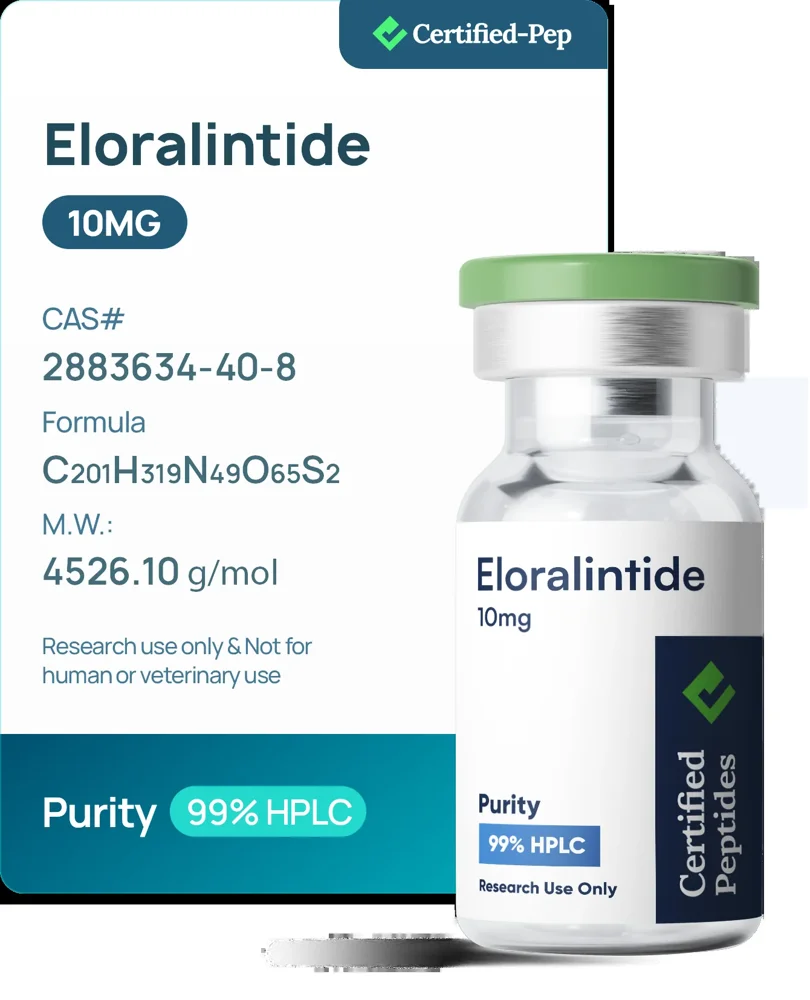
Eloralintide 10mg
$280.00USD · source price
↗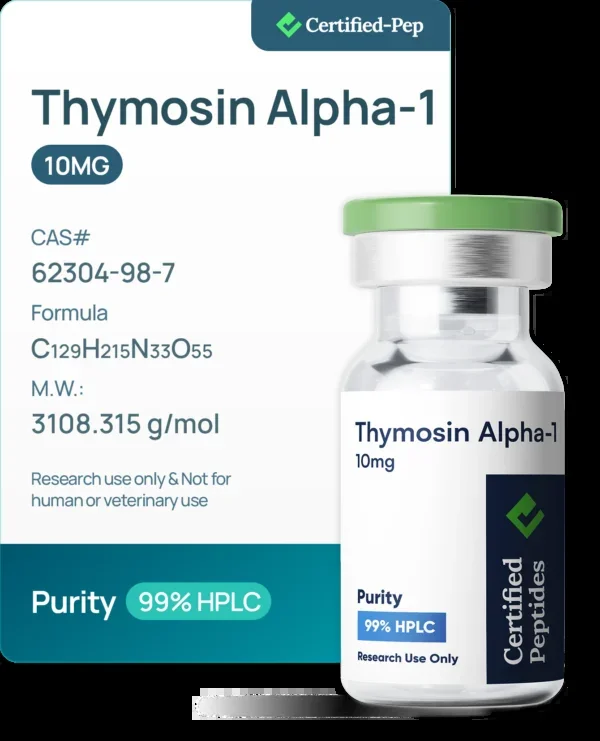
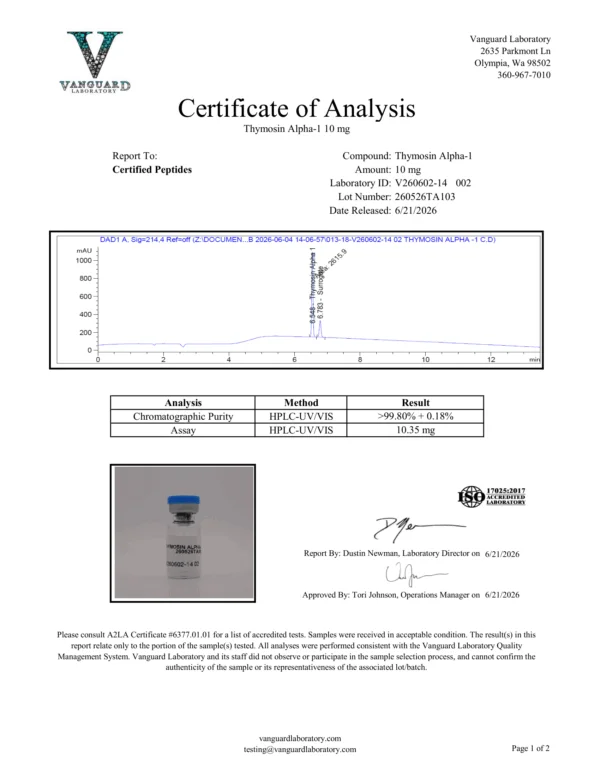
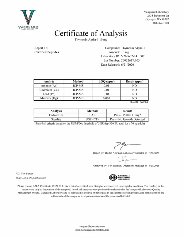
Product imagery supplied by Certified Pep. Images may include packaging or report graphics; confirm exact contents and batch records at the source.
Certified-Pep only sells U.S.-made peptides and products.
Key Features:
Made in the USA
Batch tested at a verifiable U.S. laboratory for identity, content and purity
Screened for endotoxins, heavy metals and sterility
Rigorous quality testing on every batch
All products are delivered in lyophilized powder form
For laboratory and research use only
Adds 1 item and opens the original store’s cart. Final availability is checked there.
| THYMOSIN ALPHA-1 10MG | |
|---|---|
| SKU | CP_THYMOSIN_ALPHA_1_10MG_3ML_VIAL |
| CATEGORY | Research Compound |
| CAS | 62304-98-7 |
| VIAL SIZE | 3 mL |
| FORM | LYOPHILIZED POWDER |
| MOLECULAR WEIGHT | 3108.32 g/mol |
| SEQUENCE | Ser-Asp-Ala-Ala-Val-Asp-Thr-Ser-Ser-Glu-Ile-Thr-Thr-Lys-Asp-Leu-Lys-Glu-Lys-Lys-Glu-Val-Val-Glu-Glu-Ala-Glu-Asn |
| MOLECULAR FORMULA | C129H215N33O55 |
| ITEM ID | 845 |
THYMOSIN ALPHA-1 10MG provides Thymosin alpha-1 as lyophilized powder in a 3 mL vial.
Research Context
Thymosin alpha-1 has been investigated in nonclinical models across immunology, oncology, and neuroscience. Below are summaries of nonclinical findings drawn from the supplied literature.
In Vitro CD8⁺ T-Cell Studies
Mishra et al. (2026) examined Tα1 effects on human CD8⁺ T-cells cultured under four conditions: untreated, CD3/CD28-stimulated, Tα1-treated, and combined CD3/CD28 plus Tα1. Using CFSE-based flow cytometry, the investigators measured proliferation indices and surface expression of activation markers (CD69, CD25, HLA-DR) and exhaustion markers (PD-1, TIM-3, LAG-3). The combination condition showed increased proliferation index and elevated activation-marker expression relative to individual treatments. In a chronic-stimulation model, Tα1 treatment was associated with reduced expression of PD-1, TIM-3, and LAG-3 on CD8⁺ T-cells. Cytokine secretion (IL-2, IFN-γ, TNF-α, IL-10) was measured by multiplex bead-based assay and was elevated in the combination group [1].
Dendritic Cell Signaling and Chemotherapy-Derived Apoptotic Bodies (Murine Model)
Wei et al. (2026) investigated Tα1 interactions with apoptotic bodies (ABs) released from chemotherapy-treated tumor cells in murine models. The study reported that Tα1 bound to tumor ABs and interacted with AB-borne miR-146a-5p within the endolysosomal compartment of dendritic cells (DCs). This interaction was associated with reduced lysosomal RNase A-mediated degradation of miR-146a-5p, facilitating TLR7 activation. Downstream readouts included DC maturation markers, migration to tumor-draining lymph nodes, and CD8⁺ T-cell activation assays. The authors described TLR7-dependent effects in tumor-bearing mice with high miR-146a-5p expression [2].
HCRTR1 Engagement in Murine Ischemic Stroke Model
Kang et al. (2026) reported that Tα1 acts as a ligand for Hypocretin (Orexin) Receptor 1 (HCRTR1) on neurons. In murine ischemic stroke models, genetic deletion of the Tα1-encoding gene Ptma was associated with increased injury markers, while Tα1 administration was associated with reduced RIPK3-mediated necroptosis signaling. The study measured circulating Tα1 levels, neuronal cell-death markers, and behavioral endpoints in mice [3].